Seeing Inside
No knives. No blood. No sticking out your tongue and saying, “Ahhhhh.” Magnetic Resonance Imaging (MRI) lets doctors look inside you safely and painlessly.
It’s a uniquely detailed diagnostic tool, sensitive enough to detect a ripple of fluid in the brain or the minute narrowing of a blood vessel - without using X-rays, which can be harmful.
Impact
Your Inner Self
Some software changes lives. MRI software saves lives.
MRI is part of a long line of non-invasive imaging tools, beginning with X-rays in 1895. But unlike X-rays or CT scans, MRI doesn’t expose patients to ionizing radiation and reveals different body structures. This lets doctors decide safely how to treat patients, reducing unnecessary procedures, unnecessary side effects…and unnecessary costs.
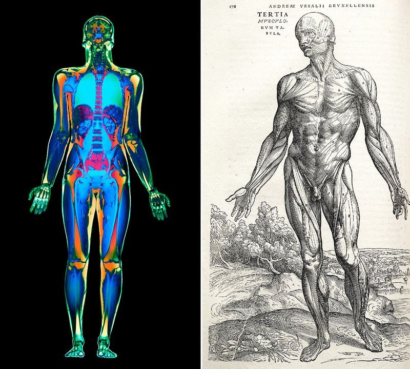
Left: Simon Fraser/Science Photo Library Right: Credit: Andreas Vesalius (public domain), via Wikimedia Commons
Left-MRI whole body scan Right-Andreas Vesalius, Di humani corporis fabrica, 1543
For centuries human anatomy remained a mystery. Public dissections and experiments were common, but yielded limited knowledge. Today, noninvasive medical imaging techniques, like MRI, enable doctors to learn what’s happening inside our bodies without cutting into them.
MRI at Work
A blood clot in the brain landed Fred S. in the emergency room. Five hours had passed, outside the usual window for using clot-dissolving drugs. Yet an MRI showed that the drugs might still work.
Fred was soon discharged —and his life saved—thanks to treatment that would not have been performed without the MRI.

Photo by Ken Glaser via Media Bakery
Brain MRIs
MRI captures 3-D pictures of an area, like your brain, by imaging different layers, called anatomical planes. The three basic anatomical planes are axial (top to bottom), coronal (front to back), and sagittal (left to right). Different abnormalities appear more clearly on different planes.
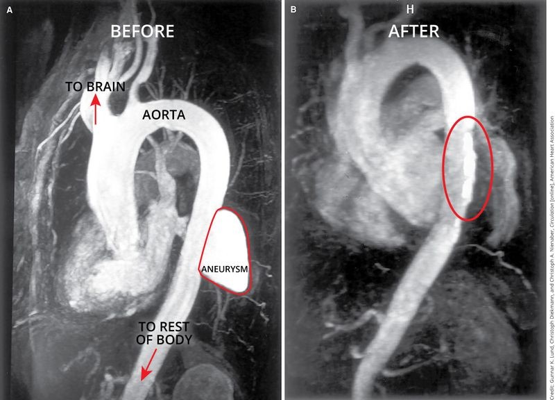
Credit: Gunnar K. Lund, Christoph Diekmann, and Christoph A. Nienaber, Circulation [online], American Heart Association
Aorta before (left) and after (right) survery
Aneurysms occur when the walls of a blood vessel become weak, causing them to bulge. Untreated, aneurysms can be fatal. These MRI images show a patient’s aorta before and after repair of an aneurysm.
© Siemens Healthineers 2016. Used with permission.
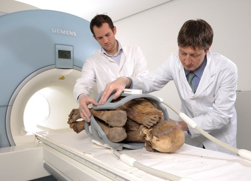
Researchers at the University of Zurich, February 11, 2008
Ultrashort echo time (UTE) is a technique that enables doctors using MRI to image materials with little or no water, like bone and cartilage, using MRI. In 2008 scientists tested UTE on a 1,000-year-old Peruvian mummy.
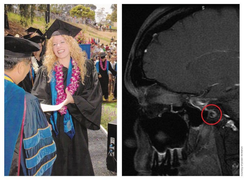
Jordan McMahon, 2005 (left) and clear MRI scan, 2013 (right)

MRI Software Makers and Users

Mobile MRI scanner
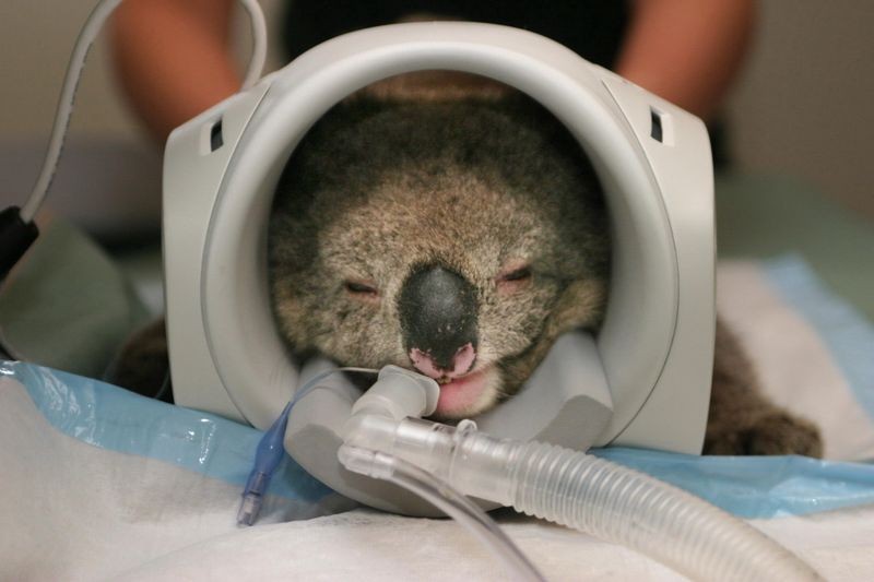
Kiki the Koala, Melbourne Veterinary Referral Centre, November 8, 2005
For Better and For Worse
Is more information always better?
MRI has transformed medicine, letting doctors look inside us before picking up a scalpel, prescribing drugs, planning treatment, and following-up.
But MRI also spots harmless abnormalities that might never cause problems, leading to tests and treatments that can be costly or dangerous—and unnecessary.

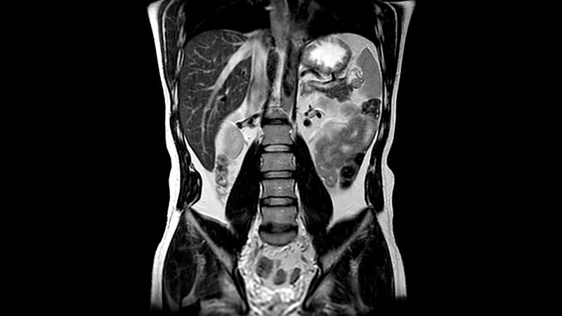
Credit: Eun Hui Bae, Young-Hwan Hwang, and Soo Wan Kim, International Braz J Urol [online]
Kidney MRI, postoperative axial (left) and coronal (right) scans
Incidental findings are abnormalities found on an MRI image while looking for something else. This liver study revealed cysts—fluid-filled sacs—in the right kidney.
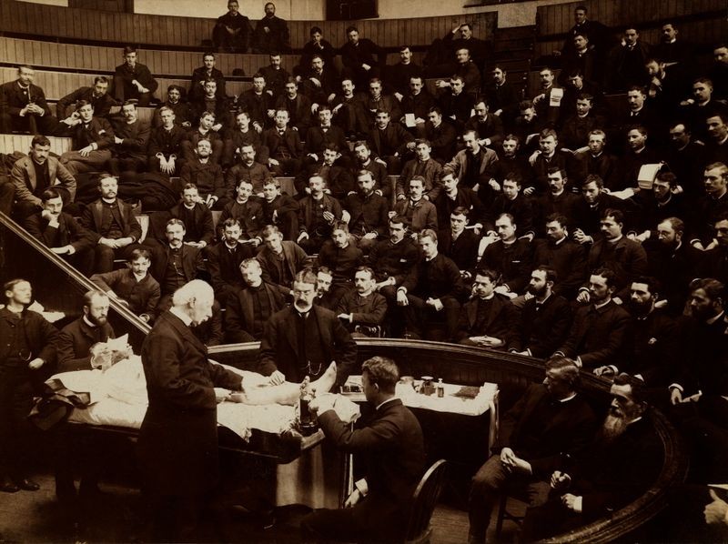
Agnew Clinic, University of Pennsylvania, March 30, 1886

Anatomage Virtual Dissection Touch Table
Technology
What is an MRI?
MRI machines use a magnetic field, radio waves, and a computer to detect the properties of living tissue. All body parts have water (and thus hydrogen).
An MRI machine creates a magnetic field around your body, causing your hydrogen atoms, which are partially magnetic, to align with the field. When radio waves are applied quickly, they excite these atoms, causing them to emit a unique electronic signal that sensors record—and which MRI software uses to create an image.
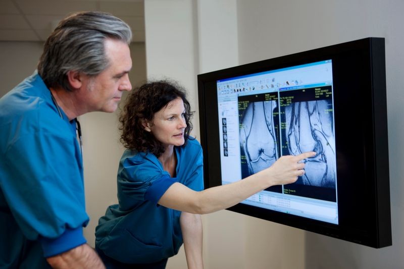
T.T. via Getty Images
Doctors evaluate knee MRI
MRI is an invaluable noninvasive diagnostic tool that combines hardware and software. Its technology produces detailed anatomical images that helps doctors detect disease, spot abnormalities, and monitor treatment.
Painting the Picture
We think of MRI “machines” as hardware. Yet much of their power lies in their software, which controls the magnetic and radio-frequency pulses, collects and interprets the resulting signals, and then transforms this data into images.
Depending on the MRI settings, images can highlight fat, water fluid, air, bone, or soft tissues in the patient.

Shutterstock/BlueRingMedia
MRI machine
MRI machines contain a large magnet that aligns the hydrogen atoms in your body. When excited by a radio pulse, those atoms emit signals, measured by radio frequency coils. Gradient coils add position information to the signal. The loud noise MRI machines make is the vibration of the gradient coils, caused by rapidly changing electromagnetic fields.
Credit: GE Healthcare
Abdominal MRI, showing fluid, fat and air
MRI detects different tissue types and displays them in varying shades of grey. This abdominal MRI scan shows fluid, fat, and air as well as major organs like the liver and lungs.
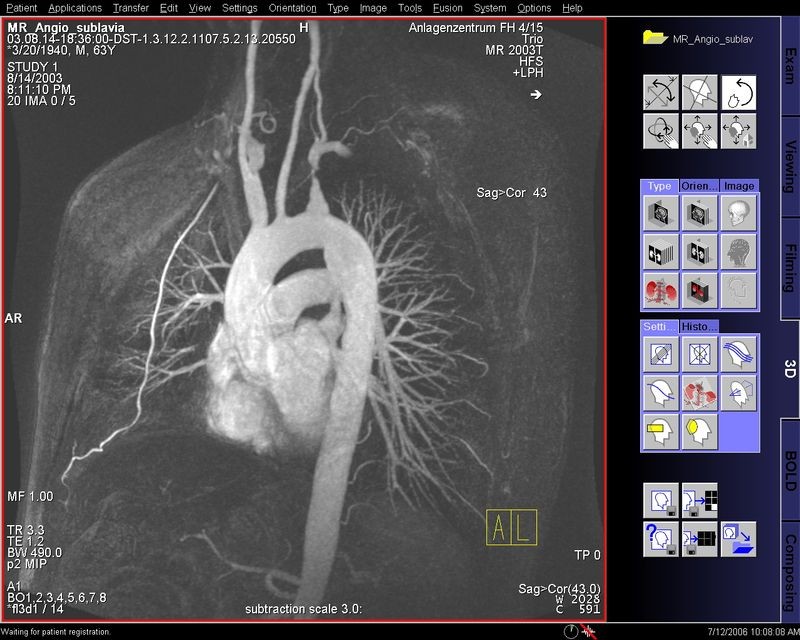
Gadolinium, a special contrast agent that illuminates blood vessels, may be given before an MRI to highlight certain tissues, like this heart and its major blood vessels.
Technician console screenshot, showing contrast agent
Credit: stereofx.org/volume
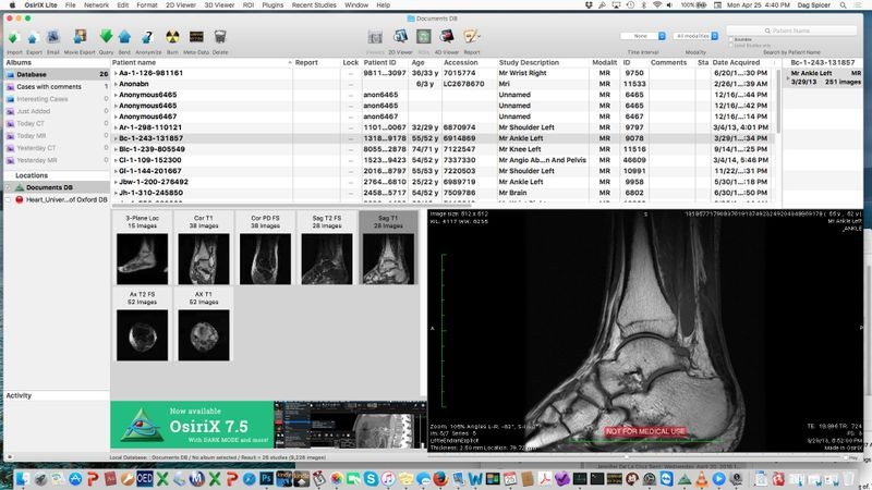
OsiriX DICOM image viewer

Eric Olcott, MD, Professor of Radiology, Stanford University School of Medicine
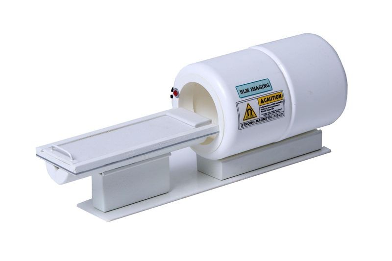
1/12th-scale model of MRI machine
Show & Tell
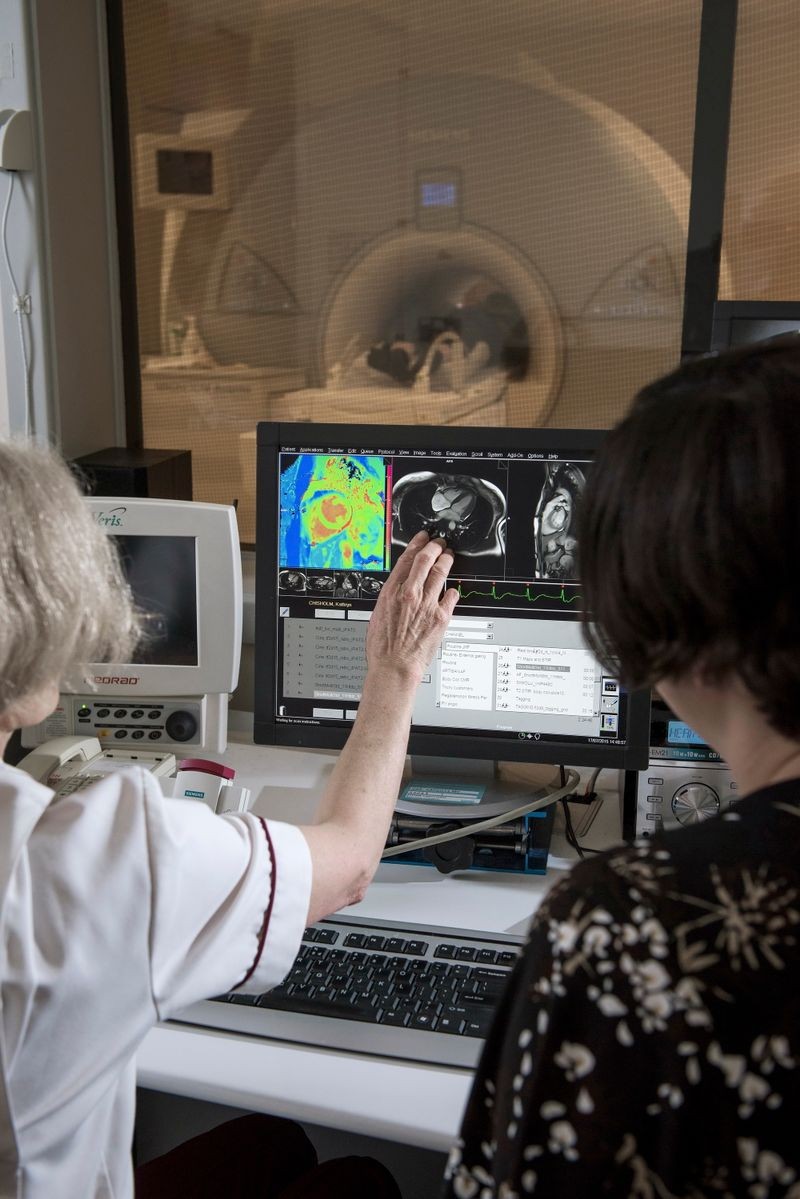
Software is a valuable tool throughout the MRI process. During your MRI, technicians use software to select imaging parameters and administer pulse sequences. After your MRI, software is used to process your images so that doctors can interpret them.
Software systems also archive and display MRI images, allowing doctors to consult with and connect patients to far-flung medical specialists when needed.
John Cairns Photography/Oxford University Images/Science Photo Library
Technician console, Oxford Centre for Magnetic Resonance, John Radcliffe Hospital, December 1, 2015
Technicians ensure MRI machinery is running properly and verify that the correct pulse sequences are used. They monitor patients from a separate room, but can communicate with patients via a two-way intercom.

Surgeon examines MRI data sets
History
Creating MRI Technology
MRI evolved over decades, beginning in 1937 with Isidor Rabi’s discovery of nuclear magnetic resonance (NMR).
Knowing that an atom’s protons and neutrons act as small, spinning magnets, Rabi exposed different compounds to a large magnetic field and powerful radio waves. Each gave off a unique signal—a magnetic signature.
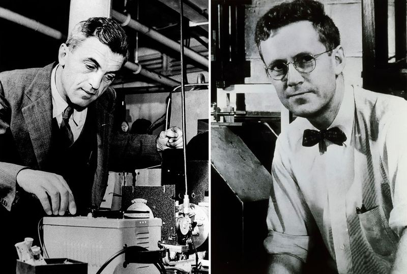
A decade later, Felix Bloch and Edward Purcell built on Rabi’s work, independently demonstrating NMR in liquids and solids.
Emilio Segre Visual Archives/American Institute of Physics/Science Photo Library
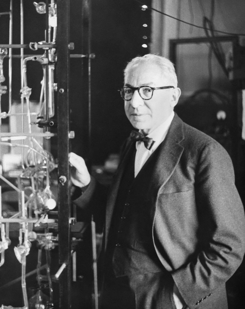
Isidor Isaac Rabi
Isidor Isaac Rabi was awarded the 1944 Nobel Prize in Physics for his method for recording the magnetic properties of atomic nuclei. It became the foundation for MRI and nuclear magnetic resonance spectroscopy.
Data to Images: An Evolution of MRI
Collecting molecular data was the first step in developing MRI. But how to transform this data into pictures?
In the 1970s, Paul Lauterbur and Peter Mansfield independently suggested applying precise variations in a second, “gradient,” magnetic field to pinpoint resonating molecules. Using this information, MRI software assembles a 2- or 3-dimensional image of internal body structures.

NYPL/Science Source via Getty Images
1930s: Isidor Isaac Rabi, 1944 Nobel Prize
Development of molecular beam magnetic resonance by passing a beam of lithium molecules through a magnetic field and then bombarding the beam with radio waves.
Left: Keystone-France\Gamma-Rapho via Getty Images Right: Science Photo Gallery
1940s: Felix Bloch (left) and Edward Purcell (right), 1952 Nobel Prize (shared)
Independent demonstration of nuclear magnetic resonance in liquids and solids and development of new methods for measuring magnetization of molecules.
AIP Emilio Segre Visual Archives, Physics Today Collection
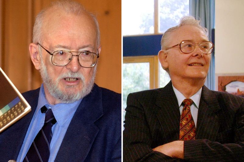
1950s: Erwin Hahn
Discovery of the spin-echo phenomenon for nuclear magnetic resonance measurements. Basic principle used in NMR and MRI today.
AIP Emilio Segre Visual Archives, Physics Today Collection
1970s: Allan M. Cormack (left) and Godfrey N. Hounsfield (right), 1979 Nobel Prize (shared)
Development of the CT scanner that uses reconstruction from projections, the foundation of nearly every many imaging systems used today.
1970s: Raymond Damadian
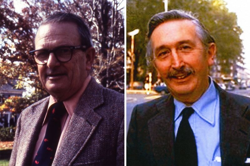
1970s: Paul C. Lauterbur (left) and Sir Peter Mansfield (right), 2003 Nobel Prize (shared)
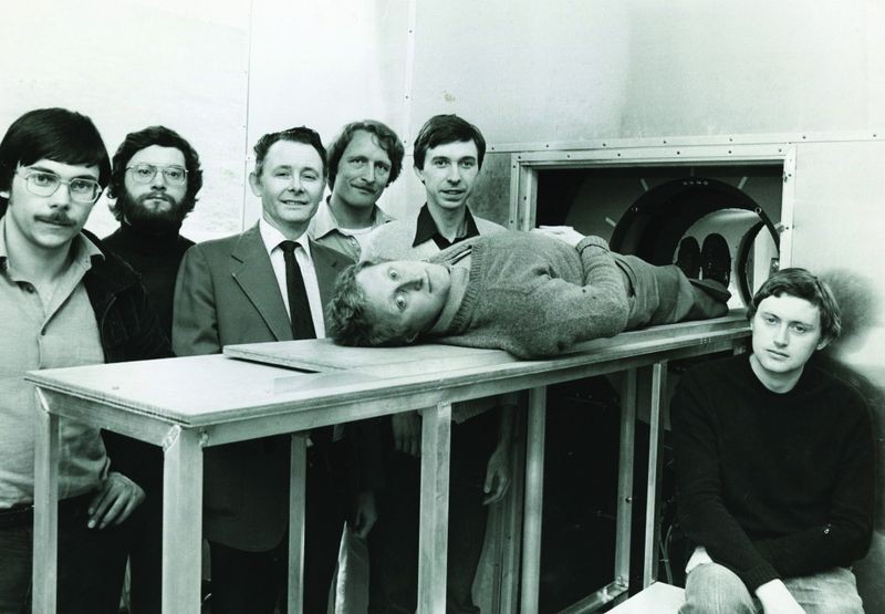
1980s: Whole-body MRI team
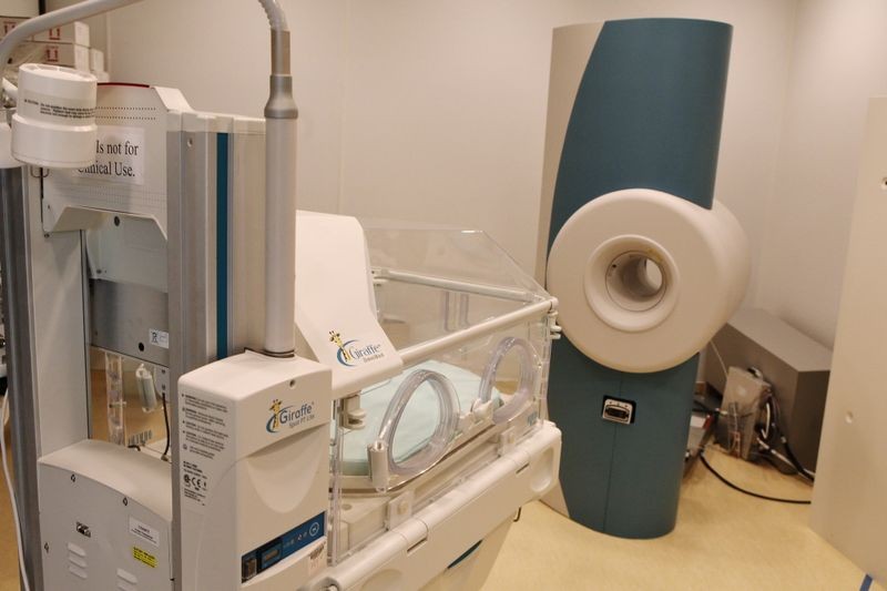
2013: Neonatal MRI machine

2015: Open MRI